Digital imaging for light microscopy
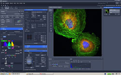
The ZEN 2012 software makes Carl Zeiss microscopes and cameras complete, transforming them into powerful, customized imaging stations. With little training you can control the entire workflow of your laboratory applications interactively.
Depending on the camera, you will be able to acquire individual images, multichannel fluorescence images or video sequences with up to 16 bits of image information per channel. ZEN provides you with reliable support by always suggesting the optimum dye and wavelength combinations for an experiment.
After acquisition you will also find important camera and device information in the very place you need it. Our specially developed image format documents the entire history of your image for this purpose. These image meta data enable you to reproduce acquisition situations and underline your results.
Do you just want to acquire microscope images and perform simple image processing or do you require ZEN together with one of our software modules? With three different versions of the software, you are guaranteed to have the functionality at your disposal to meet all the requirements of everyday laboratory practice.
Get to know the new Carl Zeiss microscope software today free of charge and without obligation with our new entry-level version ZEN lite 2012.
Digital Image Processing Software for your microscope
Whether you are a beginner in digital microscopy or an imaging expert, AxioVision will guide you to reproducible results with the aide of structured workflows. With our digital image processing software, you control essential parts of your Carl Zeiss imaging system. For example the objective turret, digital cameras, motorized stages, or fast-switching filter wheels. All of the microscope settings and processing steps are adjusted quickly and easily in a single user interface.
Only with our software you will enjoy the benefits of the Carl Zeiss ZVI image. This file format was developed specifically for the needs of scientific microscopy. Digital images are processed and saved uncompressed or with a loss-free wavelet algorithm. In contrast to generic file formats like TIF or JPEG, additional data about your experiment is stored with the image. The ZVI file contains basic information of microscope parts used for acquisition, like for example the objective. In addition to that you will be informed of the exact time points images were captured during the course of a multi-day experiment. You can also annotate comments, processing steps or measurement data to your image.
AxioVision allows to visualize and present your images in several dimensions. The functionality of this imaging toolbox expands constantly with a wide range of different modules that are tailored to specific applications or microscope accessories. Now and in the future, you need only one microscope software for your lab - AxioVision from Carl Zeiss.
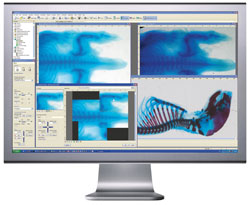
Based on over 25 years of input from Image-Pro Plus software users, Image-Pro Premier offers intuitive tools that make it easy to capture, process, measure, analyze and share your images and valuable data. The new Image-Pro Premier offers 64-bit support, a user-friendly interface, intuitive macros and app building tools, new and improved ways to automatically segment, classify and measure objects, and more tools for customizing your workflow.
 Create, Download and Share Custom Apps -
Image-Pro Premier Apps allow you to easily design step-by-step workflows that walk you through your image analysis processes. Download apps from the Solutions Zone website or choose to develop and package your own apps to share with colleagues.
Create, Download and Share Custom Apps -
Image-Pro Premier Apps allow you to easily design step-by-step workflows that walk you through your image analysis processes. Download apps from the Solutions Zone website or choose to develop and package your own apps to share with colleagues.
 Capture Single Images and Movies - Successful analysis begins with high quality images. Analyze live images, and stream multi-gigabyte movies directly to your hard drive.
 Process and Enhance -
Reveal important details within your images with filters, color adjustment tools, alignment and tiling features.
Process and Enhance -
Reveal important details within your images with filters, color adjustment tools, alignment and tiling features.
 Measure Distances and Areas, Track Objects, and Measure Intensity
- Extract quantifiable data from your images with a variety of measurement tools.
Measure Distances and Areas, Track Objects, and Measure Intensity
- Extract quantifiable data from your images with a variety of measurement tools.
 Automatically Count and Classify Objects
- Count and characterize objects using over fifty manual and automatic measurement tools. Automatically find dark or bright objects or use Smart Segmentation to threshold difficult images.
Automatically Count and Classify Objects
- Count and characterize objects using over fifty manual and automatic measurement tools. Automatically find dark or bright objects or use Smart Segmentation to threshold difficult images. Automate Your Tasks - Automation tools not only save time by eliminating repetitive steps, but more importantly, they minimize the chance of errors or inconsistencies.
 Share Your Work - Now that your analysis is complete, you need to share your findings. Annotate, export to Excel , send images to PowerPoint, create PDF reports and share an audit trail of your imaging steps.
 Designed for Your Application - Image-Pro software is used worldwide by thousands of researchers and imaging professionals in a wide range of applications including life science research, pathology, fluorescence imaging, ring analysis and aging, cell biology, industrial inspection, quality control, particle analysis, forensics and more.
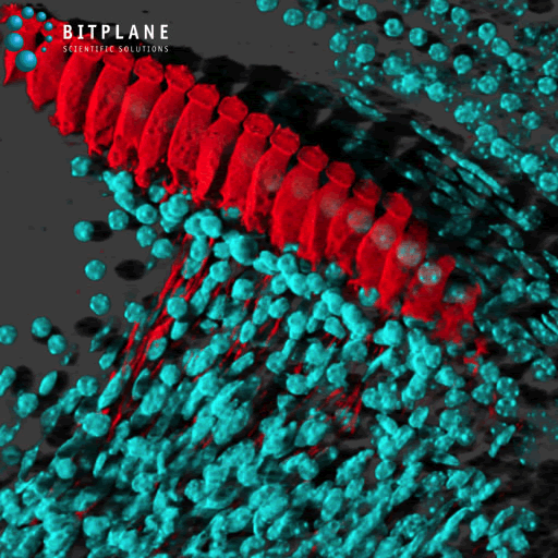
Imaris is Bitplane’s core scientific software module that delivers all the necessary functionality for data visualization, analysis, segmentation and interpretation of 3D and 4D microscopy datasets. Combining speed, precision and ease-of-use, Imaris provides a complete set of features for working with three- and four-dimensional multi-channel images of any size, from a few megabytes to multiple gigabytes in size. Conveniently load, process and visualize data and images acquired from almost any confocal and wide field microscope to gain new and groundbreaking insight from your image data. Imaris uses Standard Scientific Notation throughout.
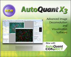
Advanced Image Deconvolution and 3D Visualization Software for Life Science Researchers.
AutoQuant is the life science industry’s leading image deconvolution software. Retrieve better data from your images using the most complete suite of 2D and 3D restoration algorithms available, including the industry’s best blind deconvolution algorithm. Microscopy experts worldwide trust AutoQuant for the accuracy and beauty of its stunning quantitative results, while newcomers to the product love the user-friendly workflow and intuitive interface that helps make learning a breeze.
• AutoQuant Connect Workflow Accelerator – Send to AutoQuant with a single mouse-click!
• All new High Resolution Interface
• Custom Gibson-Lanni Point Spread Function (PSF) Modeling
• Auto Detect Unidentified Sample Dimensions
• Sync Multiple Line Profiles and Measurements for comparison
• ROI Deconvolution Preview
• Deconvolution and Optical settings files now saved in .cfg format
• Faster Deconvolution Times
• Faster loading of images into the Batch Processing queue
• New additions to the Objective, Camera, and Dye Lists
• Much more!
Compare AutoQuantX3 to Previous Versions
Quickly compare the latest features of the AutoQuant X3 with features found in previous versions. See how AutoQuant X3 can work best for you and how it supports your existing hardware and specific applications.
Features
AutoQuant offers the most complete suite of 2D and 3D deconvolution algorithms available. Choose to save time and money using the industry's best blind algorithms that calculate Point Spread Functions (PSF) without having to spend countless hours capturing images of beads or opt to supply measured PSF images for AutoQuant to process related volumes quickly and easily. AutoQuant makes it simple to prepare image sets quickly, or import one of the many supported proprietary file formats, stabilize image sets with alignment tools, deconvolve image sets in 2D or 3D, visualize time, Z, and channel, and then analyze all parameters within the same advanced application.